
Simple Squamous Epithelium Definition
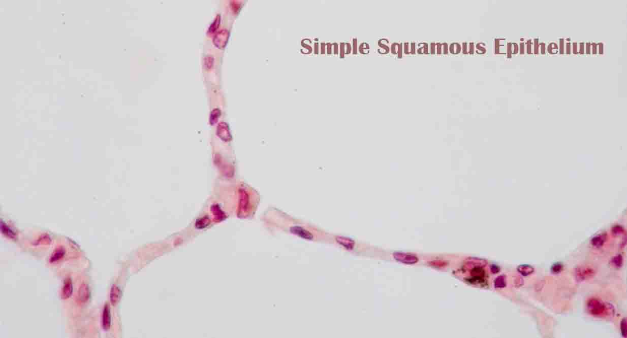
Surfaces are lined with simple squamous epithelia, which are formed from one layer of squamous cells. The nucleus of a squamous cell is rounded and large, thin, and flat. They possess polarity and a distinct apical surface with specialized membrane proteins, like other epithelial cells.
Functions of Simple Squamous Epithelium
These epithelia are commonly found in areas where absorption or transport of materials are important. Additionally, they are involved in diffusion, osmosis, and filtration. In the kidney, the alveoli of the lungs, and the walls of capillaries, they play a significant role.
It is an ideal medium for selective transmembrane transport since it is composed of a single layer of tightly packed thin cells. There are some substances that travel mainly along their concentration gradients, such as oxygen from the lungs. Proteins bound to membranes are used for active transport of others.
Tight junctions are formed by membrane proteins to ensure that ions, gases, small molecules, and water are transported only through the cell and not through the interstitial spaces. Basolateral surface is composed of the rest of the plasma membrane.
Examples of Simple Squamous Epithelia
From capillaries to alveoli in the lungs, and nephrons in the kidneys, simple squamous epithelium can be found in a variety of places. The majority of these cells originate in the embryo’s ectoderm, or outermost layer. There are, however, some simple squamous epithelial tissues derived from the mesoderm or middle layer of embryonic cells.
Particularly in pathological studies, they are called mesothelia. They line several body cavities, including the pleural cavity where the lungs reside, the peritoneum (the abdominal cavity where the stomach and intestines are located) and the mediastinum (part of the thoracic cavity where the heart, lungs, trachea, and esophagus are located). When they form the inner lining of blood vessels and lymphatic vessels, these tissues are called endothelium.
Example I: Alveoli of the Lung
Upon entering the body, air passes through pharynxes, larynxes, tracheas, and the branching network of bronchi to end in alveoli. A pulmonary alveoli is similar to a set of florets at the end of a stalk. Squamous cells make up this highly vascularized bulbous structure that is surrounded by capillaries.
Gas is exchanged between alveoli and blood vessels through capillaries, which are made up of a single layer of squamous cells. About 12,000 liters of air pass through the lungs each day, supplying oxygen to the body and expelling carbon dioxide. This important bodily function is mediated by the two sets of simple squamous epithelia.
Example II: Bowman’s Capsule in the Kidney
Nutrients and wastes are transported through the blood by this organ. As a result of protein and nucleic acid metabolism, nitrogenous substances are one of the most important waste products of the body.
Structures called glomeruli in the kidney remove these substances from the blood. There are one million functional subunits called nephrons in the kidney. It is the Bowman’s capsule at the end of a nephron that interacts closely with a glomerulus.
Bowman’s capsule surrounds the capillaries, which are red in color. Bowman’s capsule is composed of squamous cells on the outer surface and modified squamous epithelium on the inner surface, near capillaries. Podocytes are specialized cells that form slit-like structures on the inner surface of the viscera to facilitate filtration.
Each day, nearly 180 liters of blood are filtered between the squamous epithelial cells in the glomerular capillaries and those in the Bowman’s capsule. During the course of a day, each blood cell passes through this structure about 20-25 times.
Through a series of secretions, absorptions and reabsorptions, urine is eventually formed and expelled from the body from the glomerular filtrate at the start of the nephron.
Related Biology Terms
- In epithelial cells, the apical surface faces the organ lumen or the external environment.
- The basement membrane is a supportive structure formed by extracellular matrix secreted by epithelial cells.
- It is a thin membrane made of modified carbohydrates and proteins that separates epithelial cells from connective tissue.
- Specifically formed connections between cells, especially in the epithelium, that are formed by close interaction between two sets of transmembrane proteins that prevent materials from moving between cells.